Animals
 Animation
 Art of Playing Cards
 Drugs
Drugs Education
 Environment
 Flying
 History
 Humour
 Immigration
 Info/Tech
 Intellectual/Entertaining
 Lifestyles
 Men
Men Money/Politics/Law
 New Jersey
 Odds and Oddities
 Older & Under
 Photography
 Prisons
 Relationships
 Science
 Social/Cultural
 Terrorism
 Wellington
 Working
 Zero Return Investment
Where Is Consciousness? I've Lost It!
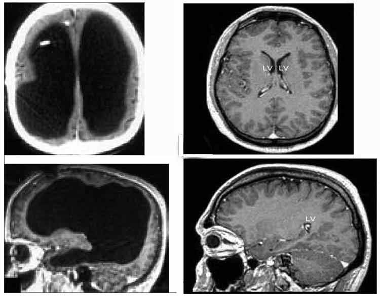
Is the Brain Really Necessary?Aristotle taught that the brain exists merely to cool the blood and is not involved in the process of thinking. - Will Cuppy This was the question asked by British neurologist John Lorber when he addressed a conference of pædiatricians in 1980. Such a frivolous sounding question was sparked by case studies Lorber had been involved in since the mid-60s. The case studies involve victims of an ailment known as hydrocephalus, more commonly known as water on the brain. The condition results from an abnormal build up of cerebrospinal fluid and can cause severe retardation and death if not treated. Two young children with hydrocephalus referred to Lorber presented with normal mental development for their age. In both children, there was no evidence of a cerebral cortex. One of the children died at age 3 months, the second at 12 months. He was still following a normal development profile with the exception of the apparent lack of cerebral tissue shown by repeated medical testing. An account of the children was published in Developmental Medicine and Child Neurology. Later, a colleague at Sheffield University became aware of a young man with a larger than normal head. He was referred to Lorber even though it had not caused him any difficulty. Although the boy had an IQ of 126 and had a first class honours degree in mathematics, he had "virtually no brain". A noninvasive measurement of radio density known as CAT scan showed the boy's skull was lined with a thin layer of brain cells to a millimeter in thickness. The rest of his skull was filled with cerebrospinal fluid. The young man continues a normal life with the exception of his knowledge that he has no brain. Although anecdotal accounts may be found in medical literature, Lorber is the first to provide a systematic study of such cases. He has documented over 600 scans of people with hydrocephalus and has broken them into four groups:
Of the last group, which comprised less than 10% of the study, half were profoundly retarded. The remaining half had IQs greater than 100. Skeptics have claimed that it was an error of interpretation of the scans themselves. Lorber himself admits that reading a CAT scan can be tricky. He also has said that he would not make such a claim without evidence. In answer to attacks that he has not precisely quantified the amount of brain tissue missing, he added, "I can't say whether the mathematics student has a brain weighing 50 grams or 150 grams, but it is clear that it is nowhere near the normal 1.5 kilograms." Many neurologists feel that this is a tribute to the brain's redundancy and its ability to reassign functions. Others, however, are not so sure. Patrick Wall, professor of anatomy at University College, London states "To talk of redundancy is a cop-out to get around something you don't understand." Norman Geschwind, a neurologist at Boston's Beth Israel Hospital agrees: "Certainly the brain has a remarkable capacity for reassigning functions following trauma, but you can usually pick up some kind of deficit with the right tests, even after apparently full recovery." ReferencesAnthony Smith The Mind New York Viking Press, 1984, page 230 Roger Lewin "Is Your Brain Really Necessary?"Science 210 December 1980, page 1232 KeelyNet BBS (214) 324-3501 There are no restrictions on duplicating, publishing or distributing the files on KeelyNet except where noted! Source: web.syr.edu 30 October 1993
Dev Med Child Neurol. 1992 Jul; 34(7):623 - 32. Related Articles, Links Reciprocal neurological developments of twins discordant for hydrocephalus. Berker E, Goldstein G, Lorber J, Priestley B, Smith A Studies of 10 sets of twins discordant for hydrocephalus in early life revealed striking differences in degree and nature of development of verbal versus non-verbal cognitive functions, birth order, and hand and eye preference. Despite similar (4 dizygotic pairs) or identical (6 monozygotic pairs) genetic endowment and grossly similar intra- and extra-uterine environmental and socio-economic influences, the consistency of the differences between the hydrocephalic children and their seemingly normal twins indicate systematic differences in pre-, peri- and/or early postnatal organisation and development of hemispheric function. Follow-up studies also documented development of above-average intelligence, despite drastically reduced cerebral mantle size in hydrocephalus of early onset. The atypical patterns of development of the non-hydrocephalic twins also confirm previously described qualifications reported in studies of the significance of genetic cersus environmental factors in twins. Publication Types: Source: ncbi.nlm.nih.gov
More about Roger Lewin's Science article on Lorber's claims and his research posted on the blog Metafilter.com: "...As a rule, material is not published in Science if it is not credible, for the magazine is one of the flagship publications of the English speaking scientific world - so reputations are at stake... Lorber has his supporters and detractors in the medical community. But, as the Science article notes, the medical literature is replete with anecdotal accounts:
Experimental research on cats cited in the article seem to support some of Lober's claims. I would also note that Lorber studied hydrocephalic conditions probably more extensively than anyone in the world, before or since. I think that what really underlies the whole controversy - and what probably drove Lorber to employ some hyperbole in scandalizing the medical community with his "virtually no detectable brain" statements or his stock "Is your brain really necessary" lectures - was the tendency, in the medical community and elsewhere, to marginalize anomalous observed phenomenon, to shove such troubling stuff into a closet where it can be safely ignored. There was a whole slew of such anomalous material, preceding probably even the extensive observations of Goethe, which was ignored for years until it was exhumed by mathematicians such as Mandelbrot; these observed anomalies led mathematicians along the trail to the development of chaos theory (or the mathematics of nonlinear equations, to use a less charged term for the now sprawling field). Paul Pietsch, at University of Indiana, has done quite a bit of research into another such anomalous, fascinating (or infuriating) phenomenon. Oddly, the sarcastic conjecture I made about the "squished" brains "inflating" when holes are drilled in hydrocephalic patient's heads are correct ... sort of (this happens with the installation of a shunt, see below). The below text is from Roger Lewin's Science story on Lorber's claims. Lorber himself acknowledges that the "Virtually no brain" claim was hyperbolic: "As to the question "Is your brain really necessary?" Lorber admits that it is only half serious. "You have to be dramatic in order to make people listen." So Lorber used the tongue-in-cheek hyperbole to shock the medical community. He didn't intend it as a strict scientific claim. Lorber came to make his observations on hydrocephalus through his involvement with assessment and treatment of spina bifida, a congenital condition in which the spinal column fails to fuse completely, leaving nerve tissue perilously exposed. The great majority of patients with spina bifida also suffer from hydrocephalus. Although the origins of hydrocephalus are to some degree shrouded in mystery, it is clearly associated with a disturbance of the circulation of cerebrospinal fluid through a system of channels and reservoirs, or ventricles, in the brain. Back pressure apparently develops, and this may balloon the ventricles to many times their normal size, so pressing the overlying brain tissue against the cranium. In young children, whose skulls are still malleable, one obvious consequence can be a grossly enlarged head. Additionally, this physical assault from within leads to a real loss of brain matter. It is therefore not surprising that many hydrocephalics suffer intellectual and physical disabilities. What is surprising, however, is that a substantial proportion of patients appear lo escape functional impairment in spite of grossly abnormal brain structure. "The spina bifida unit at the Children's Hospital here in Sheffield is one of the largest in the world," explains Lorber, "and this gives us an opportunity to make many observations. Since the introduction of the safe, noninvasive brain scanning technique just a few years ago we have done more than 600 scans on patients with hydrocephalus." Lorber divides the subjects into four categories :those with minimally enlarged ventricles; those whose ventricles fill 50 to 70% of the cranium; those in which the ventricles fill between 70 to 90% of the intracranial space; and the most severe group, in which ventricle expansion fills 95% of the cranium. Many of the individuals in this last group, which forms just less than 10% of the total sample, are severely disabled, but half of them have IQ's greater than 100. This group provides some of the most dramatic examples of apparently normal function against all odds. Commenting on Lorber's work, Kenneth Till, a former neurosurgeon at the Great Ormond Street Hospital for Sick Children, London, has this to say: "Interpreting brain scans can be very tricky. There can be a great deal more brain tissue in the cranium than is immediately apparent." Till echoes the cautions of many practitioners when he says, "Lorber may be being rather overdramatic when he says that someone has 'virtually no brain.' "Lorber acknowledges the problem of interpretation of brain scans, and he counters Till's remarks by insisting, "Of course these results are dramatic, but they're not overdramatic. One would not make the claim if one did not have the evidence." A major obstacle in this work is the difficulty of obtaining the kind of quantitative data that would be expected in a scientific investigation of, say. rat brains. "I can't say whether the mathematics student has a brain weighing 50 grams or 150 grams, but it's clear that it is nowhere near the normal 1.5 kilograms,"asserts Lorber, "and much of the brain he does have is in the more primitive deep structures that are relatively spared in hydrocephalus." Lorber concludes from these observations that "there must be a tremendous amount of redundancy or spare capacity in the brain, just as there is with kidney and liver." He also contends that "the cortex probably is responsible for a great deal less than most people imagine." These are two areas of considerable dispute in neurobiology. Wall lends support for this second point. "One reason why results such as Lorber's have been neglected for so long is because of the implied attack on the predominance of the cerebral cortex,"suggests Wall. "For hundreds of years neurologists have assumed that all that is dear to them is performed by the cortex, but it may well be that the deep structures in the brain carry out many of the functions assumed to be the sole province of the cortex." He likens the cortex to a "reference library"that may be consulted from time to time. Norman Geschwind, a neurologist at the Beth Israel Hospital, Boston, strikes a different note. "Deep structures in the brain are undoubtedly important for many functions,"he agrees, "but I don't believe the explanation that the cortex does far less than we think is very sound." And neither does David Bowsher, professor of neurophysiology at Liverpool University, England: "I don't think we attribute more to the cortex than it deserves." Bower, however, takes the middle ground, with the suggestion that "the deep structures are almost certainly more important than is currently thought." On the question of the brain's spare capacity there is equal contention. "To talk of redundancy in the brain is an intellectual cop-out to try to get round something you don't understand,"states Wall. Geschwind agrees: "Certainly the brain has a remarkable capacity for reassigning functions following trauma, but you can usually pick up some kind of deficit with the right tests, even after apparently full recovery." However, Colin Blakemore. professor of physiology at Oxford University, England, sees spare capacity as an important quality of the human brain. "The brain frequently has to cope with minor lesions and it's crucial that it can overcome these readily,"he says, "there may be some reorganisation of brain tissue, but mostly there's a re-allocation of function." It is perhaps significant that many of the instances in which gross enlargement of cerebral ventricles is compatible with normal life are cases where the condition develops slowly. Gross surgical lesions in rat brains are known to inflict severe functional disruption, but if the same damage is done bit by bit over a long period of time, the dysfunction can be minimal. Just as the rat brains appear to cope with a stepwise reduction of available hardware, so too do the human brains in some cases of hydrocephalus. Another subgroup of some curiosity in Lorber's subjects are those people in whom expansion of the ventricles is restricted to just one side of the brain. "I've now seen more than 50 cases of asymmetric hydrocephalus,"says Lorber, "and the interesting thing is that only at minority of these individuals show the expected and long-cherished neurological finding of paralysis with spasticity on the opposite side of the body." To make matters even more puzzling, one individual in the group has enormously enlarged ventricles on the same side as his spastic paralysis. "This is exactly the opposite to all that we learnt in medical school,"reports Lorber with obvious glee. These observations are cogent support for Bower's comment that "the concept of contalateral control is the least secure of all our concepts about brain organization and function." Lorber's extensive series of brain scans stands in marked contrast with the dearth of information on the fine structure of hydrocephalic human brains. "It is crucial to know about the histological state of the brains of these functionally normal hydrocephalic patients,"remarks Lorber, "but how am I to have access to such material, given the ethical barriers to scientific research on patients?" Inadequate though it is, the next best thing is experimental work on animals. A group of researchers based at the New York University Medical Center has assembled a picture of the histological changes associated with hydrocephalus through experimental induction of the condition in cats. The group also observed the changes in tissue structure following the implantation of a shunt, the experimental equivalent to the normal treatment of hydrocephalus in humans. Speaking for the group, Fred Epstein says the following: "Hydrocephalus is principally a disease of the white matter. As the ventricles enlarge the layers of fibres above them begin to be stretched and very quickly they are disrupted, with the axons and the myelin sheaths surrounding them breaking down. Even in severe and extended hydrocephalus, however, the nerve cells in the gray matter were remarkably spared, though eventually there began to be a loss here too." The sparing of the gray matter even in severe hydrocephalus could go some way to explaining the remarkable retention of many normal functions in severely affected individuals. Crucial to the approach to treatment of hydrocephalus is the brain's ability to recuperate following the release of fluid pressure when a shunt is implanted. One of the canons of neurobiology is that, once damaged, cells in the central nervous system are unable to repair themselves. Does Lorber's work dent this hallowed concept too? "When you implant a shunt in a young hydrocephalic child you often see complete restoration of overall brain structure, even in cases where initially there is no detectable mantle,"claims Lorber. "There must be true regeneration of brain substance in some sense, but I'm not necessarily saying that nerve cells regenerate,"he says cautiously; "I don't think anyone knows fully about that." What, then, is happening when a hydrocephalic brain rebounds from being a thin layer lining a fluid-filled cranium to become an apparently normal structure when released from hydrostatic pressure? According to Epstein and on the basis of his colleagues' observations on experimental cats, the term rebound aptly describes the reconstitution process, with stretched fibres shortening, thus diminishing the previously expanded ventricular space. Within a short time scar tissue forms, constructed from the glial cells that pack between the nerve cells. "The reconstitution of the mantle,"report Epstein and his colleagues, "does not result in the reformation of lost elements, but rather in the formation of aglial scar and possibly a return to function of the remaining elements." Lorber claims that his observations on the dramatic recovery of severely affected young children imply that "clinicians shouldn't give up in the face of an apparently hopeless case; a shunt operation at an early stage has a good chance of producing a normal individual." In mild cases, or ones that develop slowly and late, Lorber takes a different approach. Citing the example of the mathematics student and others like him, he proposes that perhaps the surgical knife should be stayed, "because a shunt operation makes an individual forever dependent on surgical care, and in any case many of these subjects can lead perfectly normal lives." The difference is between the acute and chronic conditions. These statements are certain not lo go unchallenged, partly because there is a multiplicity of opinions about appropriate treatment of hydrocephalus and partly because it is Lorber who is making them. Lorber is no stranger to controversy. Just a few years ago he caused a storm in the medical world by suggesting that it is not always medically right to administer extensive treatment to some infants with spina bifida. His experience had taught him that the consequences in some severe cases were simply not tolerable, either to the patient or to the immediate family. This position continues to be hotly debated, but Lorber's ideas are beginning to receive favourable consideration, particularly in the United Kingdom . What of the Lorber approach to hydrocephalus? "His attitude is based on many years of clinical experience,"says Gerald Hochwald of New York University Medical Center, "and it contains a certain amount of value." Thomas Milhorat, a neurosurgeon at the Children's Hospital in Washington, DC, voices strong support for Lorber, in spite of many differences of opinion. "I'm glad there's a John Lorber,"says Milhorat; "he could be more moderate in the way he expresses things, but a moderate view would not emerge if someone were not speaking out strongly." As to the question "Is your brain really necessary?"Lorber admits that it is only half serious. "You have to be dramatic in order to make people listen,"concedes the tactician. Bower's answer to the tongue-in-cheek question is this: "Although Lorber's work doesn't demonstrate that we don't need a brain, it does show that the brain can work in conditions we would have thought impossible." Bower occasionally complains that Lorber's style is less scientific than it might be. He concedes, however, that "there are still many questions to be answered about the human brain, and it has to be admitted that Lorber's provocative approach does make you think about them." Source: metafilter.com posted by troutfishing 29 June 2004
Tiny Brain No Obstacle to French Civil Servant
The large black space shows the fluid that replaced much of the patient’s brain (left). Washington - A man with an unusually tiny brain managed to live an entirely normal life despite his condition, caused by a fluid buildup in his skull, French researchers reported. Scans of the 44-year-old man's brain showed that a huge fluid-filled chamber called a ventricle took up most of the room in his skull, leaving little more than a thin sheet of actual brain tissue. "He was a married father of 2 children, and worked as a civil servant," Dr Lionel Feuillet and colleagues at the Universite de la Mediterranee in Marseille wrote in a letter to the The Lancet medical journal. The man went to a hospital after he had mild weakness in his left leg. When Feuillet's staff took his medical history, they learned he had had a shunt inserted into his head to drain away hydrocephalus - water on the brain - as an infant. The shunt was removed when he was 14. So the researchers did a computed tomography (CT) scan and another type of scan called magnetic resonance imaging (MRI). They were astonished to see "massive enlargement" of the lateral ventricles - usually tiny chambers that hold the cerebrospinal fluid that cushions the brain. Intelligence tests showed the man had an IQ of 75, below the average score of 100 but not considered mentally retarded or disabled, either. "What I find amazing to this day is how the brain can deal with something which you think should not be compatible with life," commented Dr Max Muenke, a paediatric brain defect specialist at the National Human Genome Research Institute. "If something happens very slowly over quite some time, maybe over decades, the different parts of the brain take up functions that would normally be done by the part that is pushed to the side," he added. Muenke was not involved in the case. Source: stuff.co.nz 22 July 2007
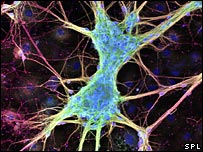
Single Brain Cell's Power Shown
Individual cells may be more powerful than thought There could be enough computing ability in just one brain cell to allow humans and animals to feel, a study suggests. The brain has 100 billion neurons but scientists had thought they needed to join forces in larger networks to produce thoughts and sensations. The Dutch and German study, published in Nature, found that stimulating just one rat neuron could deliver the sensation of touch. One UK expert said this was the first time this had been measured in mammals. The complexity of the human brain and how it stores countless thoughts, sensations and memories are still not fully understood. Researchers believe connections between individual neurons, forming networks of at least 1,000, are the key to some of its processing power. However, in some creatures with simpler nervous systems, such as flies, a single neuron can play a more significant role. The latest research suggests this may also be true in "higher" animals. The team, from the Humboldt University in Germany and the Erasmus Medical Centre in the Netherlands, stimulated single neurons in rats and found this was enough to trigger a behavioural response when their whiskers were touched. A second research project from the US suggests the computational ability of the brain cell could be even more complex, with different synapses - the many junctions between neurons and other nerve cells - able to act independently from those found elsewhere on the same cell. This could mean that, within a single neuron, different synapses could be storing or processing completely different bits of information. Dr Douglas Armstrong, the deputy director of the Edinburgh Centre for Bioinformatics, said the research did not mean all neurons had an individual role to play but that, in some instances, they might be capable of working alone with measurable results. He said: "The generally accepted model was that networks or arrays make decisions and that the influence of a single neuron is smaller - but this work and other recent studies support a more important role for the individual neuron. These studies drive down the level at which relevant computation is happening in the brain." Source: news.bbc.co.uk 22 December 2007
What and Where Is Consciousness?Science tells us that particular areas of the brain carry out specific functions, so how do people with damaged or underdeveloped brains still function normally? In 1996 in the US, a young boy, here referred to as James, was about to undergo a serious operation. James was only eight years old and suffered from a condition known as Sturge-Weber syndrome, which had caused the formation of abnormal blood vessels in the left hemisphere of his brain. As a result he was afflicted by regular epileptic fits and had a very low mental age; the only word in James's vocabulary was "Mamma." In an attempt to to rectify the problem, doctors felt forced to take drastic steps. They decided to remove the entire left side of his brain the medical team knew that, since the left side of the brain controls the right side of the body, the operation to save James's life would also leave him partially paralysed. What they didn't expect, however, were the developments in James's condition which occurred soon after the surgery. Within weeks, James began to talk and, two years later, was close to reaching a normal mental age. Amazingly, the operation to remove an entire hemisphere of his brain appears to have cured him of his learning difficulties. Such remarkable examples of adaptability are far more common than we might think. In conflict with established medical thinking, there are literally hundreds of cases where people have either been born with an underdeveloped brain, or have had large areas of their brain damaged in an accident, but are still able to function normally. Such anomalies were partly explained when it was discovered that we have the ability to relocate particular brain functions to other areas of the brain. Exactly how this works is still beyond modern science, and so the ability lies in limbo between accepted medical fact and that which is still regarded as nonsense. However, it may be that this discovery is only the tip of the iceberg, for there are people whose very existence seems to indicate that our brains are nowhere near as vital to our survival as we might think. In countries across the world, there are hundreds of cases of a condition called hydrocephalus (often known colloquially as "water on the brain"), where cavities form in the brain that can be so large that they account for 95% of the brain's mass. This leaves only a fluid-filled bubble of the outermost cerebral tissue which, in extreme cases, has been found to be less than one millimetre thick. (Ordinarily, the walls of the cerebrum are 45mm thick.) The condition is so serious that, if it is recognised before birth, a decision is often taken to terminate the pregnancy because only a small proportion of sufferers survive. In those born with this condition, the body's production of the cerebrospinal fluid (CSF) which fills the cavities in the brain is working at a rate well above the norm. This usually leads to a swelling of the cranium; one six-year-old boy had a skull with a circumference 72cm greater than that of the average adult. Modern techniques, however, allowed doctors to drain the fluid until normal pressure was restored and the boy survived. Despite the seriousness of this condition, in some people it appears to have little or no affect on their intellectual abilities. Indeed, to the surprise of the medical establishment, in a study of 253 hydrocephalus sufferers carried out by the University of Sheffield, Professor John Lorber discovered that there is no relation between volume of brain tissue and IQ. Surprising ResultsOf the 253 subjects in the study, 9 were found to have approximately only 5% of the normal amount of brain tissue. Despite this, 4 had IQ's of above 100, the national average, and another 2 had IQ's of above 126, while one of the subjects proved to be as intelligent as those studying him, he had a first-class degree in maths. One possible explanation for such achievements as this is the neopallium, which forms the very outermost layer of the brain. Since the brains are larger with hydrocephalus sufferers, they have larger neopalliums while the brain mass is diminished in bulk. The neopallium is the site for some of the most important mental functions, such as the power of reasoning. Cases such as these have been cropping up regularly to test the stability of modern medicine, yet are largely disregarded. They undermine established beliefs about the relationship between the human brain and the site of consciousness and so are largely ignored by mainstream medical science. When asked about the impact of his research into hydrocephalus sufferers, Professor Lorber said it had "suffered a fate like much of the literature of phenomenological science: it was ignored." While science chooses to blinker itself, these medical anomalies continue to walk the streets, their fluid-filled craniums not preventing them from leading normal lives and taking degrees. Brainless BoyOne related case that has received more exposure than most is that of Andrew Vandal, who was born on 12 July 1984. In the early stages of his development in the womb a cyst appeared on the stem of his brain. Known as an atelencephic aprosencephaly, this destructive event left the boy with a cranium containing nothing but fluid. In some cases, it can even leave victims with no detectible brain at all - a condition known as anencephaly or "brainlessness." Cases like Andrew's are again usually terminated before birth, but in this instance the subject was born and then put up for adoption. He was adopted by a pædiatric nurse, Kaye Vandal, from Wallingford, Connecticut, US, who, when last asked about Andrew's welfare, stated that she remained devoted to "giving him the best quality life for however long he lives." At the same time, Kaye stated that, against doctors' predictions, Andrew was able to laugh, giggle and smile and, has a "glowing, outgoing, bubbly personality." Kaye also stated that her young charge was able to respond to stimulus and was maturing mentally; both of which doctors believed to be impossible, considering his complete absence of brain matter. Andrew was, however, unable to speak, and was cortically blind; that is, he could see images, but was unable to interpret them. Andrew was also incapable of walking, but did manage to drag himself along on his back. Cases such as Andrew's provide real-life testimony to our astonishing adaptability as biological organisms. Source: mysteries.pwp.blueyonder.co.uk
For articles on bacteria, centrioles, chairs, nebulae, asteroids, robots, memory, chirality, pain, fractals, DNA, geology, strange facts, extra dimensions, spare parts, discoveries, ageing and more
click the "Up" button below to take you to the Index for this Science section. |