Animals
 Animation
 Art of Playing Cards
 Drugs
Drugs Education
 Environment
 Flying
 History
 Humour
 Immigration
 Info/Tech
 Intellectual/Entertaining
 Lifestyles
 Men
Men Money/Politics/Law
 New Jersey
 Odds and Oddities
 Older & Under
 Photography
 Prisons
 Relationships
 Science
 Social/Cultural
 Terrorism
 Wellington
 Working
 Zero Return Investment
Expiration Date
Loving Rover to DeathDachshunds are ideal dogs for small children, as they are already stretched and pulled to such a length that the child cannot do much harm one way or the other. - Robert Benchley Ever consider what pets must think of us? I mean, here we come back from a grocery store with the most amazing haul - chicken, pork, half a cow. - Anne Tyler Brief Livesby David Jones "A dog is for life - not just for Christmas!" This slogan is intended to warn potential purchasers of the long-term consequences of pet ownership: not merely dogs, and not merely at Christmas. Yet thousands of households buy pets to amuse the children, and are stuck with them when the children are no longer amused. Now Daedalus has the answer. He is inventing the short-life pet. In principle, no mammal can live forever. At every cell division, one unit is lost from the telomere sequence on each of its chromosomes. When the entire telomere sequence has gone, the cell can no longer divide. Its vital reserve is exhausted. So Daedelus plans to shorten the telomere sequence in pet animals. His biologists are taking fertilised dog, cat and hamster ova, and cultivating them in vitro by standard methods. Every animal, of course arises from a single fertilised ovum. It divides into two cells, then Into four, and ultimately into a complete fœtus. If at the two-cell stage the cells are separated, they go on to develop into identical twins. The DREADCO team members are encouraging this process. Each time an ovum divides into two, they separate the two cells. When these reach the two-cell stage, these are separated again, and so on. At each cell-division, one telomere unit is lost from the cells' chromosome termination. After many such divisions, Daedalus will have millions of identical telomere-depleted ova of the chosen pet species. They will be implanted into surrogate mothers of the same species, and brought to term. The resulting identical youngsters will command vast prices in the pet market. Children will adore their fluffy appeal. Canny parents will appreciate their brevity. Almost as soon as their appeal has waned, their telomere sequences will be exhausted, and they will drop dead. Daedalus is not sure what they will die of. It won't be senility; brain cells hardly divide at all. Gut cells divide very fast, so it may be indigestion. But one standard feature of old age, in Man as in animals, is the increasing embrlttlement and fragility of connective tissue. So their fibroblast teleomeres may give out first. Like other toys, Daedalus's short-Iife pets may just fall to pieces. Source: Nature Vol 404 2 March 2000
I have to ask if this would be sending the proper message to children. Short-life parents may be next. I think teaching children that there are long-term consequences to short-term pleasures that must always be taken into account may be a better, and more realistic, message.
Dying for a SteakExpiry Date Guaranteedby David Jones Last week Daedalus invented the short-life pet. By repeatedly dividing a newly fertilised ovum in vitro, he obtained large numbers of single ova, each of which had already undergone many cell divisions. Every time a cell divides, each of its chromosomes loses a telomere unit from its termination; when they have all gone, it can divide no longer, and is doomed. So Daedalus's telomere-depleted ova have a limited life ahead of them. Brought to term in a surrogate mother, they are sure to die young making them perfect pets. This process has further implications. Do even human identical twins, who have undergone one more cell division than the rest of us, have a slightly reduced lifespan? Probably not. Few of us die from telomere exhaustion; indeed, even Daedalus finds it hard to guess how it would show itself. With luck it will be a mercifully swift extinction, as one whole class of body cells - in the gut, say, or in the liver - suddenly ceases to replicate. If so, Daedalus sees applications in humane animal husbandry. At present, sadly, all farmed animals have to be deliberately killed. Any animal that dies a "natural death" has probably succumbed to some pathogen and its meat is unsafe to eat. But an animal that dies of telomere exhaustion would be entirely safe. With proper timing, it could be arranged to drop dead just after having reached its ideal size and condition. Even better, repeated ovum division creates unlimited numbers of such animals, all identical. Implanted into surrogate mothers, they would all have the same predictable lifespan. DREADCO agronomists are now studying the logistics of a humane agriculture in which identical ova are implanted at regular intervals into production line of surrogate mothers. The animals are brought to term and allowed to grow up normally. On the predicted day of their death, they are transported to the slaughterhouse by modern "just in time" delivery methods. They walk onto the conveyor belt in the correct order, and each drops dead just as it reaches the butchery section. No violence is necessary. Animal-rights enthusiasts will cheer, consumers will welcome the reliable consistency of the products, and accountants will revel in the utter predictability of it all. Source: Nature Vol 404 9 March 2000
I expect the anæsthesia discussed in the article Sedatives Reduce Stress in Salmon (earlier in this section) would be a better answer for now - although I understand other painless, non-detectable means of euthanasia are being developed. It's about time. One solution may be to get your kids pigs for pets. By the time the child gets bored of their pet, the pig will be big enough to eat. Or you could develop a taste for dog meat - see the article on the following page (press the "Next" button below). For more on problems with the relationships between animals, man and food see the articles on hunting, slaughtering, eating1, eating2, and pests (all found elsewhere in this section).
A Curious Immortality
Over the past 45 years, research on HeLa cells has provided scientists with an enormous amount of basic knowledge about the physiology and genetics of cells. However, HeLa cells have been responsible for generating a great deal of bogus scientific data as well. It turns out that HeLa cells grow very aggressively in culture and can easily invade other cell cultures during routine lab transfer procedures, when proper precautions are not taken. As a result, numerous research papers have been published on the biology of a variety of cultured cell types which have subsequently been shown to be HeLa cells. Karyotype (chromosome) analysis of HeLa cells from different repositories around the world shows that different strains of HeLa cells are now very different from each other, probably due to the malignant nature of the cells and differences in culture conditions in different laboratories over the decades since this cell culture was established. Although HeLa cells provided a substantial foundation for today's knowledge of cell physiology, most analysis of cell structure and function in culture is now performed with non-transformed (not malignant) cells. Some biologists believe that HeLa cells are no longer human at all and consider them to simply be single-celled microorganisms. Tom Cavanagh Source: www.madsci.org 7 April 1997
Helacyton gartleri: The Immortal Remains of Henrietta LacksWhen human body cells are removed and put into a cell culture, they weaken and die quickly, usually within about 50 divisions. Without the rest of the support structure - a heart, blood circulating, a digestive system and so-on - body cells can't survive. Body cells also age, so even if you were to simulate the body's environment in a test tube or Petri dish, the cells would eventually perish anyway. The basic mortality of the cells reflect the basic mortality of the organism they comprise, which is why there's no fountain of youth or medicinal procedure that'll give you biological immortality. There is, however, one human being who is biologically immortal on a technicality, and her name is Henrietta Lacks. In 1951 she showed up at John Hopkins Hospital in Baltimore, complaining of blood spotting in her underwear. Samples were taken of her cervical tissue and sent to a lab for analysis, which came back with a diagnosis of cervical cancer. The cancer was caused by the human papillomavirus, which is a sexually transmitted disease. Most variants of this virus are harmless, but some are known to cause cervical cancer, as in Henrietta's case. After her diagnosis and before attempts to treat the disease with radium, another sample from the tumour was sent to George Gey, who was the head of tissue culture research at Hopkins. Gey discovered that the cells from Henrietta's tumour would not only survive and multiply outside of her body, but they didn't age either. These cells were basically immortal. They're still alive. Now, HeLa cells are about as common in biological research as the lab rat and the Petri dish, and are still being grown in an unbroken lineage from the cells originally harvested from Mrs Lacks in 1951. They're used in cancer research because a scientist can perform experiments on them that otherwise couldn't be done on a living human being. They were also used in the development of the polio vaccine, making Henrietta posthumous hero to millions. Say you're a scientist looking at HeLa cells under a microscope. They live independently of the body they came from. They reproduce (faster even than other cancerous cells). They consume, excrete, and do everything an independent living organism usually does. A thousand years from now there will still be HeLa cells multiplying and living, even some original cells sampled from Mrs Lacks, though Henrietta herself has long since passed away. Is this a new species? In 1991 the scientific community decided it was, and blessed HeLa cells with its own genus and species: Helacyton gartleri. Helacyton gartleri is an example of speciation, which is when a new species is observed developing from another. In this case, the development is from a chordate (homo sapien) to something that's more like an amœba (a cross-phylum mutation), giving us an animal with a mostly human genotype, but which does not develop into a human-like phenotype. Since this event occurred in nature when the papillomavirus transformed Henrietta's cells, and not in the laboratory, it's a strong piece of evidence supporting evolution (although not one that suggests you could go from an amœba to a chordate, which would probably take more than one mutation). Source: www.disenchanted.com
The HeLa Cell, a Biological First for African America!In biological and medical research, a HeLa cell is a cell which is derived from cervical cancer cells taken from an African American woman named Henrietta Lacks. The cervical cancer cells from Ms Lacks were circulated (without her knowledge or permission) by George Gey. These cancer cells are considered "immortal" (that is, they do not die of old age and can divide an unlimited number of times, unlike most other human cells), and have been grown in cell culture in an unbroken lineage ever since. This cell line was propagated for use in cancer research. Initially, the cell line was said to be named after a "Helen Lane", in order to preserve Lacks's anonymity. The cells proliferate abnormally rapidly, even compared to other cancers. HeLa cells have been transformed by human papilloma virus 18 (HPV18), and have different properties from normal cervical cells. They are used as model cancer cells and for studying cellular signal transudation. HeLa cells have proven difficult to control. They sometimes contaminate other cell cultures growing in the same laboratory, interfering with biological research. The degree of contamination is unknown, because few researchers test the identity or purity of already-established cell lines. It has been claimed that a substantial fraction of in vitro cell lines are actually HeLa, their original cells having been overwhelmed by a rapidly growing population derived from HeLa contaminant cells. It has been estimated that the total mass of HeLa cells far exceeds that of the rest of Henrietta Lacks' body. Researchers argued that these cells are a separate species because they reproduce and spread on their own so in 1991 it was named Helacyton gartleri. On 4 October 1951, Henrietta Lacks died of cervical cancer at Johns Hopkins University Hospital in Baltimore. But Lacks will live forever in laboratories and research centres worldwide that use her unique, immortal cells for medical research. The cells of her cancer, known as HeLa cells, were the first human cells discovered to thrive and multiply outside the body, seemingly forever, allowing researchers to conduct experiments previously impossible. Reference:The Johns Hopkins Magazine April 2000 Source: www.aaregistry.com 1 October 2004
Impostor Cells Are Wrecking Medical Researchby Andy Coghlan Countless research projects around the world into cancer and other major diseases are producing bogus or misleading results because investigators are studying the wrong type of cell. The mistakes arise when fast-growing "rogue cells" contaminate cell cultures and swamp the correct cells. A team's work on prostate cancer, for example, might turn out to be worthless because cells researchers thought were prostate cancer cells turn out to be cervical cancer cells. Although the danger has been known about for decades, most researchers still fail to check the identity of the cells they are working with. And several new types of rogue cell are emerging. Warnings of the potential scale of the problem were issued this week at a conference in Warwick organised by the UK's Health Protection Agency. Precise figures are hard to come by because researchers are either unaware that they have worked with the wrong cells, or try to cover it up. "If people have spent 3 years working on the wrong cells, they are not likely to want to tell people about it," says David Lewis, manager of the European Collection of Cell Cultures, based at Porton Down in Wiltshire. He will emphasise the importance of getting cells from authenticated sources at this week's conference. The best estimates available suggest around a fifth of all experiments in fields such as cancer and microbiology involve the wrong cells. "Various figures between 20 and 40% have been aired," says Rod MacLeod, head of the genetics laboratory at the DSMZ, the German collection of cell cultures in Braunschweig. In a study in 1999, MacLeod found that of 252 cancer cell lines, 18% were "impostors". And newly established cell lines were just as likely to be contaminated as older ones. He blames this on the ease with which cell lines can be contaminated. The most notorious rogue cells are so-called "HeLa" cells. They rapidly overrun more sluggish colonies. "Just one HeLa cell could survive and proliferate," MacLeod says. Attempts to purge cell lines contaminated by HeLa cells have waxed and waned since 1967, when the problem first emerged, but now there are new rogue cells to deal with too. One posing particular problems in cancer research is the T24 line of bladder cancer cells. MacLeod and Hans Drexler, director of the human and animal cell collection at the DSMZ, discovered them in 2002 "posing" as healthy epithelial cells that line organs. They are now popping up all over the place, especially in cultures of other cancer cells. Recently they contaminated prostate cancer cell lines, as Adrie van Bokhoven of the University of Colorado Health Sciences Center in Denver discovered. "Two years after our research, there are still publications that describe it as prostate," van Bokhoven says. In a review to be published in November 2003 in The Prostate Journal, van Bokhoven will highlight the problems posed by another rogue cell, an unusually lively prostate cancer cell line called PC-3. He reveals that another 3 "unique" prostate lines were actually PC-3, adding to others he has already discovered. Before journals publish a paper van Bokhoven wants editors to insist that researchers provide proof that their cell lines are authentic. "Editors should demand it," says van Bokhoven, who points out that the DNA fingerprinting tools now available should allow the problem to be eradicated. "There are no more excuses," he says. John Masters of University College London, who has struggled for years to highlight the problem, agrees. "I've written to journal editors till I'm blue in the face on this, and they do nothing," he says. This unwillingness to confront the issue has been dubbed "false cell-line denial syndrome" by Drexler, who says that the problem will persist as long as it is swept under the carpet. Senior figures at the American Type Culture Collection have also tried for years to highlight the problem, says Keith Bostian, of the American Society of Microbiology. The ASM is aware of the problem, he says, and its journals "encourage" authors to deposit cell lines in public collections for authentication. Yet one survey conducted by the ATCC revealed that two-thirds of journals still fail to cite the source of biological materials. Worse, many of the cultures the ATCC receives for deposit are misidentified or contaminated. Source: www.newscientist.com New Scientist 19 September 2003 © Reed Business Information Ltd See also:
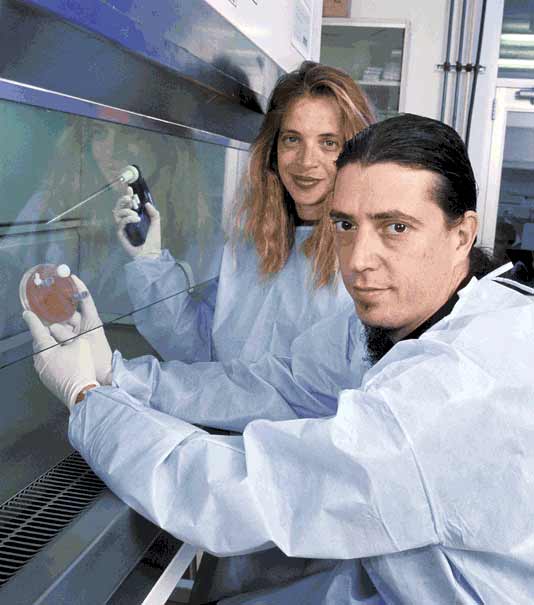
The Art of Science
Oron Catts and Ionat Zurr in the lab with their "art" work by Marit Slavin Is it possible to produce a fur coat without killing an animal? The answer, in a word, is yes. Artists Oron Catts and Ionat Zurr have done it, and the result is on view in an installation called "Victimless Leather" at the Israeli Center for Digital Art in Holon. The two grew a miniature coat from living skin cells, in an improvised laboratory they established for this purpose at the centre. The growth process, which requires a special technology, took place in the lab. For 11 days the two artists "fed" the cells, which grew and multiplied, creating a miniature coat. On the last day they "killed" the coat by no longer feeding it. The audience that viewed the display was taught how to create this kind of system by themselves. The work by Catts and Zurr is part of an exhibition titled "Free Radicals," featuring projects that reach beyond the boundaries of the arts into other fields. The result is an intriguing discourse on the limits of freedom of expression, democratization of knowledge and the question of whether it is permissible, in the name of art, to do what would be prohibited in any other sphere. Catts, 39, and Zurr, 37, a married couple with a 3-year-old daughter, are among the leaders of a relatively new field known as BioArt. Their careers and artistic development unfolded in Perth, Australia. Catts arrived there at the beginning of the 1990s, followed shortly afterward by Zurr. They worked at odd jobs and began studies at the University of Western Australia. Catts took product design and visual art; Zurr, photography and communications. "Our interest in BioArt started with a project I did in 1995 as part of a research study I had to do in product design," Catts relates. "I chose to integrate biotechnology and product design, with the aim of creating an environmentally-friendly design. At the time I thought, naively, that we had to change the conception of our consumer society to one that is more concerned about its products, instead of buying and throwing away. The idea was to use biological knowledge to create products of this kind." At the time, the media was being inundated with descriptions of a mouse that had a large human ear transplanted on its back. This experiment was conducted in the laboratories of Joseph Vacanti and Robert Langer, the fathers of tissue engineering. They created a biodegradable polymeric scaffold in the shape and size of a human ear, and it was then seeded with human cells. The polymeric scaffold gradually degraded, while concurrently, the cells grew and multiplied upon it. The final result was an artificial ear composed entirely of human cells. "For the public, this mouse symbolised the concept that it all had to do with genes, and that this was one of the dangers that lurk for us in human genetic engineering, which is a totally unfounded view," Catts says. "What we learned is that tissue engineering makes it possible to grow a tissue in 3 dimensions, not necessarily in connection with anything human. So why not grow products using this technology?" Catts continues: "We found this subject problematic. For example, if we were to take a consumer product and wrap it in living tissue, would we consider it something living? This and other issues challenged us, and we decided to go on developing the research as art, because art has the advantage of addressing paradoxes in a way that is externalized, and not necessarily coming up with solutions. The most basic definition of art is the creation of objects whose mission is to challenge the viewer so he will reconsider his worldviews, and thus to forge a cultural discourse. We tried to see whether it is possible to create objects that are constructed of living materials and present them as artistic objects." Zurr: "Art is not necessarily the creation of an object; it's enough to create a situation. At a certain level, we look at our works as performance art, in the sense that the process is more important than the finished product. The works change over time, and we document them. We are establishing the laboratory in the gallery itself, and the processes - the feeding ceremony and the ceremony of killing the cells - are carried out in front of an audience. Generally we invite the audience, the curator and the gallery owner to take part in killing the artistic object we created." Catts: "Because we are utilising tissue engineering, we can use relatively large objects in terms of cell tissues, whose average size is 4 to 5 centimeters and which can be seen with the naked eye. We are interested in people experiencing a semi-living object, and when they return to the exhibition, they will see that the object has changed. We remove the cells from an animal and 'persuade' them that the external conditions we provide them are their natural place and that they are in fact growing. We call the growing environment of the cells the techno-scientific body. At the metaphorical level, we are all living in a techno-scientific body, because we are technology-dependent organisms." Zurr: "We define ourselves as artists, researchers and curators, in the sphere of art and biology, art and life, and art and new knowledge. Tension exists between art and science, because the conception is generally of one in the service of the other, and we want to get away from that dichotomy. From our point of view, this is art that deals with new concepts of life, some of which are emerging from new concepts of the life sciences." ... Another project Catts and Zurr exhibited was called "Disembodied Cuisine." "It all started at Harvard," Catts relates. "There was a scientist there who was working on tissue engineering in the womb. She removed muscle tissues from the fœtus of a sheep, manipulated them, and returned them to the womb with the aim of seeing whether the manipulations would be manifested in the newborn. The muscle tissues multiplied like crazy in the lab and filled all the incubators. Rather than throw out the cells, the scientist offered them to staff of the lab for use. It reminded us of what our parents used to tell us, 'You don't throw away food.' We were hungry artists in Boston, and thought of food went to our head. We asked ourselves, 'Why not grow food from these muscle tissues without killing an animal?' The truth is that hypocrisy underlies this declaration, because we know that the serum that serves as food for cells that are growing and multiplying comes from animal matter, and to obtain it, it is necessary to kill lambs. The irony is that the technology distances the victim. In the lab, we grew lamb muscle cells on degradable polymers, and we obtained a steak the size of a 10-shekel coin. After 3 months of growth, the polymers disappeared completely, but the problem was that we couldn't eat the small steak, because the lab didn't have a license to serve food. "In 2000, we were the first to grow food from tissue cultures. In 2003, we were invited to a major exhibition which focused on art and biotechnology in Nantes, France. There we set up a laboratory of tissue cultures using completely new equipment, with no fear of infection. We decided to make steaks out of the most repulsive meat, and to that end used frog cells. Later, we decided to make things even more repulsive, and because we knew that the French loathe engineered food, we decided to use engineered muscle cells of frogs. From a French researcher we obtained a biopsy of engineered muscle cells from the legs of a frog. We built the laboratory and for 2 months we fed the steaks. Every day we had a ceremony of feeding the steaks. On the last day we held a big meal with the participation of 8 volunteers, in which the steak was eaten. The thing is that we ate our art, which is the most interactive thing imaginable. "We removed the steak (which was, as described, the size of a coin) from the bioreactor in which it was growing, marinated it in Calvados overnight, and cooked it in gravy that included garlic and honey. It was the ultimate nouvelle cuisine. However, the steak was not really tasty, and four people spit it out. What happened was that the polymers did not completely degrade, and because the muscle cells had not 'exercised,' they were like jelly. We quickly collected what people spat out and we are now exhibiting it in an exhibition on leftovers in disembodied cuisine. At the same time, we documented the entire process and created a video installation on 3 screens that tells the story of the project." Catts: "Not long afterward, a young scientist started to send us questions about the project, which for us was very good, because it made it possible for us to draw conclusions. We discovered that a gram of steak cost us $650, and when we calculated how much serum we had used to grow the steak, it turned out that a whole lamb would have needed to be killed for this. In other words, there are no free meals and there is no victimless situation. "The scientist who approached us wrote a long article in a respected scientific journal about the use of tissue engineering technology to grow meat substitutes, and he gave us credit. Effectively, he treated us like scientists, not artists. The story was published worldwide. Subsequently he established, in partnership with the University of Maryland, a company called New Harvest, and the government of Holland invested €5 million in a similar study. This was a very interesting development, in that artists who are engaged in a subject conceptually are suddenly showing the way. That is not what we intended, but that is what happened." Zurr: "At that time we received an e-mail from People for Ethical Treatment of Animals, a very militant American animal-welfare organisation. The organisation's leader had a project proposal: that we should take a biopsy from her and grow from her tissue a steak that she would eat. The idea was to protest the eating of animals, but this would be an act of cannibalism, which we did not like, and we refused." Catts and Zurr are preoccupied with the ethical paradox that underlies their work. "At the abstract level, technology, particularly of the Western variety, deprives us of the possibility to make ethical decisions about how we conduct our lives, because the victims are so remote that they become transparent," Catts says. "It starts with buying meat in the supermarket and proceeds to advanced war technologies. In both those cases, you don't see the victim. There is something inbuilt in the conception of Western consumerism and progress to make it possible for us not to know the price the Other pays for our way of life. This subject interests us very much, because we, too, are taking part in the representation of this false utopia. "I gave a talk at the Tate Modern in London about our work, and an art critic said he was shocked that we were capable of treating life like that. This man was wearing a full-figure leather suit. Before replying, I said to him, 'I have a problem with your clothing. At least one cow had to be slaughtered for it.' People were certain I had paid him to do it. For him, that element was so transparent that he never gave it any thought." Zurr: "On the other hand, we were in Spain and we saw that the entire Spanish society had become more American. Opposition to bullfighting had intensified, but they are eating more at McDonald's. In the wake of our work, Oron became a vegetarian, but, as he says, with conscious hypocrisy. He eats only cold-blooded creatures." Catts: "There are a great many people who are self-righteous, and moralize to others. We are conducting our lives with conscious hypocrisy; the self-righteous do the opposite. We present our works directly, so they will not preach. Our message is far more implicit - we do not provide answers. I think art loses its force when it moralizes or becomes self-righteous. The important thing for us is to bring about a situation in which those who are exposed to our work will be challenged in regard to their concept of life, and will then think about how to cope with that. All we want to tell the viewers is that the time has come to cope with it. ... Catts and Zurr are artists in residence at the School of Anatomy and Human Biology at the University of Western Australia in Perth, where they have established a laboratory that fuses art and science, called "SymbioticA." They started with a small grant, to which, in time, were added grants from artistic bodies in Australia. "We were invited to Harvard and other universities to lecture, which led to the recognition by a number of scientists in the school that the relations between science and art had to be consolidated within the framework of the university," Zurr relates. "At the same time, artists contacted us to ask if they could come to work with us. We decided to submit a request to the local lottery to build a studio for research artists in a building of the anatomy department. To our surprise, we got the money, and suddenly we had our own space. Slowly, we developed a residency program in which artists can do research, an academic program, workshops, and all kinds of things that made it possible for us to subsist." The program got underway toward the end of 2002, and artists and scientists from a broad range of disciplines are doing research in the art-science laboratories at the university. "The mandate we received from the university is to coordinate art and science," Zurr continues. "The artists work alongside scientists. All the artistic research must go through the safety and ethics commissions, like any other research. Oron lectures and manages the workshops, and I am doing a Phd and am responsible for the academic organisation of the project. Five to 10 researchers have been coming to us every year for the past 7 years. They work with DNA, with tissue cultures and other items. "In some way, we have become an entry gate for artists and intellectuals who cannot do research like this elsewhere. After they acquire working skills here, they are able to go back to their universities, enter labs, and do research. They have the beginnings of a language. Recently we got a call from Stanford University. They found our model to be ideal, and we now intend to form a partnership with them. True, we are very occupied with management, but the system is slowly arriving at a stage in which it nourishes itself. One of our remaining ambitions is to make it possible for scientists to do research in symbiotics, research that is based on sheer curiosity. It is important for us to have scientists come and do research in an artistic laboratory." Source: haaretz.com
For more on animals, including reptiles, crustaceans, arachnids, insects, fish, birds, pets, livestock, rodents, bears, primates, whales and Wellington's waterfront, click "Up"
below to take you to the Table of Contents for this Animals section. |
 In 1951 a physician removed cells from the cervix of Henrietta Lacks, a 31-year-old black
woman from Baltimore, and sent the cells to a lab to determine if they were malignant. The cells were malignant and Henrietta Lacks died eight months later from cervical
cancer. Henrietta Lacks' physician provided George and Margaret Gey of Johns Hopkins University with a sample of these cervical cancer cells. The sample of Henrietta
Lacks' cells was code-named HeLa, for the first two letters of her first and last name. The HeLa cell cultures survived and multiplied so well in culture, that they were soon
being shipped to research labs around the world for study.
In 1951 a physician removed cells from the cervix of Henrietta Lacks, a 31-year-old black
woman from Baltimore, and sent the cells to a lab to determine if they were malignant. The cells were malignant and Henrietta Lacks died eight months later from cervical
cancer. Henrietta Lacks' physician provided George and Margaret Gey of Johns Hopkins University with a sample of these cervical cancer cells. The sample of Henrietta
Lacks' cells was code-named HeLa, for the first two letters of her first and last name. The HeLa cell cultures survived and multiplied so well in culture, that they were soon
being shipped to research labs around the world for study.